The OMNICON Tumor Colony Analyzer is a powerful combination of the industry's most advanced image analyzer, a unique automatic stage and software dedicated to counting soft agar assays. The system is so sophisticated that it can distinguish colonies from biological debris, agar bubbles and other artifacts, yet it is extremely east to operate. The patented software package is ideally structured to expedite the processing of colony counting data and allows the ability to do multiple group analysis (i.e. scores colonies of various sizes and compares defined group breakdown to designated controls so as to calculate either growth-inhibition or growth-enhance values as needed).
|
Tumor Cloning Assay
Tumor Survival Assay
Stem Cell Assay
Chemotherapeutic Sensitivity
Soft Agar Assay
Clonogenic Assay
Mammalian Cell Survival Assay
|
|
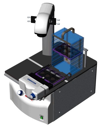
Automatic Stage
|
The OMNICON TCA system utilizes a unique automated system which combines inverted optical microscopy with a motorized X & Y stage and removable cartridge system.
Each cartridge has the capacity to transport 8 multi-well plates in varying formats (6, 12, 24, 48 and 96 wells). This versatility in footprint acceptance of cell culture plastic-ware is of critical importance since it mediates maximum flexibility to the type of colony survival assay performed.
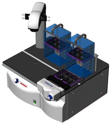
The optional Stacker Module (3 maximum) allows the system's capacity to be expanded to 32 multi-well plates. The operator can see the actual cells or colonies - not secondary reaction. Inverted optics permits the multi-well plates or petri dishes to remain covered, minimizing dehydration and contamination of the medium. Focusing is automatic for each well or dish.
|
|
With varying media and cell lines, illumination becomes an important aspect in the ability to analyze soft agar assays. The OMNICON TCA offers coaxial (transmitted and reflected) illumination with a halogen light source. For low contrast assays, the optional Phase Contrast illumination system makes those assays simple to analyze without the use of stains.
Reproducible
For research analysis, for much faster answers to vital questions, here is a tool that eliminates operator fatigue and errors. Most important, it gives repeatable results, eliminating subjective judgments and avoiding inaccuracies that occur when measuring criteria and techniques that vary between operators and laboratories.
A variety of scientific publications have been written by Doctors Mayo, Alley, Von Hoff, Herman and Hamburger which utilize the OMNICON TCA system to access potential chemotherapeutic drugs as inhibitors of malignant disease. Collectively, all the unique features demonstrated by the OMNICON TCA system make it an ideal instrument to satisfy any researcher's needs.
|
应用文献:
|
Article No.
|
Title
|
|
TCA1001
|
A Vascoactive Intestinal Peptide Antagonist Inhibits Non-small Cell Lung Cancer Growth
|
|
|
TERRY W. MOODY, FARAH ZIA, MURIEL DRAOUI, DOUGLAS E. BRENNEMAN, MATI FRIDKIN, ARIANE DAVIDSON, AND ILLANA GOZES
|

|
The most prevalent lung cancer, non-small cell lung cancer (NSCLC) has receptors for vasoactive intestinal peptide (VIP). Here the effects of a VIP antagonist (VIPhyb) on NSCLC growth were investigated. In vivo, when VIPhyb (10 µg, s.c.) was daily injected into nude mice, xenograft formation was significantly inhibited by -80%. In vitro, VIP (100 nM) stimulated colony formation -2-fold, whereas 1 µM VIPhyb inhibited colony formation by -50% when adenocarcinoma cell line NCI-H838 was used. The attenuation of tumor proliferation is receptor mediated, as VIPhyb inhibited specific 1251-labeled VIP binding to cell lines NCI-Hl57 and NCI-H838 with an IC50 of 0.7 µM. VIP (10 nM) increased the cAMP levels 5-fold when cell line NCI-H838 was used, and 10 µM VIPhyb inhibited the increase in cAMP caused by VIP. Northern blot analysis and radioimmunoassays have shown VIP mRNA and VIP-like immunoreactivity in NSCLC cells. These data suggest that VIP may be a regulatory peptide in NSCLC and that VIPhyb is a VIP receptor antagonist that inhibits proliferation.
|
|
|

|
|
TCA1002
|
Use of an Image Analysis System to Count Colonies in Stem Cell Assays of Human Tumors
|
|
|
BERNHARDT E. KRESSNER, ROGER R. A. MORTON, ALEXANDER E. MARTENS, SYDNEY E. SALMON, DANIEL D. VON HOFF, AND BARBARA SOEHNLEN
|
|
|
As is well demonstrated elsewhere in this text, the clonogenic assay for tumor colony-forming cells has applicability to a broad scope of human tumors and has proved valuable in studies of biology, clinical course, and clieinosensitivity of human cancers. The development of this promising new area of clinical research, however, has precipitated a substantial new laboratory problem-, namely, the need for automation in counting tumor colonies. This need was not fully apparent until it became clear that the clonogenic assays predicted clinical and biological features of human cancers. In the initial studies, careful qualitative and quantitative evaluations of tumor clusters and colonies in soft agar were conducted by the clinical research laboratory staff of two of the authors (S.E.S., D.D.V.H.). As their studies proceeded, we recognized that there was a major need for a precise automated instrument for selective counting of tumor colonies and therefore initiated a join developmental project with BioLogics Incorporated (formally Bausch & Lomb Incorporated) on the application of image analysis to this task.
|
|
|
|
|
TCA1003
|
Patterns Of Tumor Colony Development Over Time In Soft-Agar Culture
|
|
|
WIM J. KIRKELS, OLGA E. PELGRIM, ADRIE M. M. HOOGENBOOM, MATHILDE W. AALDERS,FRANS M. J. DFBRUYNF, G. PETER VOOUS AND CHESTER J. HERMAN
|
|
|
Human tumors were cultured by the two-layer soft-agar technique and the time course of tumor colony development was evaluated during periods of up to 6 weeks in culture. All colony counting was performed with an automated tumor colony counter (OMNICON; BioLogics Inc., Gainesville, NY, USA - formally Bausch and Lomb, Inc). This instrument provided colony counts per culture plate in six size categories from >60 Mm diameter colonies to > 149 m diameter colonies. Six to 24 culture plates were used for each "growth curve", generally 24. Control (non-drug-treated) cultures were obtained from II 7 tumors, of which 25 also provided enough cells to allow evaluation of the time course of colony development after exposure to cytostatic agents. The development of colonies in non-drug-treated plates usually demonstrated a lag phase, a logarithmic growth phase to maximum colony development and a subsequent deterioration of colonies. In spite of clumps seeded into the agar, real colony growth could be recognized by frequent colony counting of culture dishes, although the temporal patterns of growth were sometimes different if pure single-cell suspensions were compared with suspensions containing clumps from the same tumor. Drug pre-incubation caused changes in the temporal pattern of colony growth as well as in the total number of colonies. Some cultures showed drug sensitivity when evaluated at certain time points while evaluation at later time points showed only borderline drug effect or none at all. The potential utility of tumor colony growth curves in the clinical applications of tumor colony cultures is discussed.
|
|
|
|
|
TCA1004
|
In-Use Evaluation of the OMNICON Automated Tumor Colony Counter
|
|
|
CHESTER J. HERMAN, OLGA E. PELGRIM, WIM J. KIRKELS, RENE VERHEIJEN, FRANS M. J. DEBRUYNE,PETER KENEMANS, AND G. PETER VOOIJS
|
|
|
The reproducibility and accuracy of the OMNICON (BioLogics, Inc., Gainesville, VA - formally Bausch and Lomb Inc.) automated tumor colony counter for counting tumor colonies growing in double layer soft agar is evaluated and the reproducibility is compared with manual tumor colony counting. Replicate within day run-to-run colony counts of the OMNICON show a median correlation coefficient (r) of >0.985, and day-to-day median r of >0.980. In contrast, for manual colony counting, the best intra-observer reproducibility achieved is a r of 0.943 and the best inter-observer reproducibility is a r of 0.831. Analysis of results from individual culture plates counted by the OMNICON on 5 separate days shows a median coefficient of variation of 10% with 77% of the culture dishes showing coefficients of variation of colony counts over 5 days of less than 20%. Counting of culture plates during incubation shows that the OMNICON is counting tumor colonies developing after plating of a single cell suspension.
|
|
|
|
|
TCA1005
|
Improved Detection Of Drug Cytotoxicity In The Soft Agar Colony Formation Assay Through Use Of A Metabolizable Tetrazolium Salt
|
|
|
MICHAEL C. ALLEY, CINDY B. UHL, AND MICHAEL M. LIEBER
|
|
|
Use of a metabolizable tetrazolium salt was observed to facilitate assessments of tumor cell drug sensitivity in the soft-agar colony formation assay. Enzyme-mediated staining permits discrimination between viable and non-viable groups of cells so that drug-induced cytotoxicity is clearly identifiable by visual inspection as well as by computerized image analysis. The technique appears to be especially useful in the evaluation of primary tumor cell cultures which often contain substantial numbers of non-viable cellular aggregates.
|
|
|
|
|
TCA1006
|
Feasibility of Drug Screening with Panels of Human Tumor Cell Lines Using a Microculture Tetrazolium Assay
|
|
|
MICHAEL C. ALLEY, DOMINIC A. SCUDIERO, ANNE MONKS, MIRIAM L. I-LURSEY, MACIEJ J. CZERWINSKI, DONALD L. FINE, BETTY J. ABBOTT, JOSEPH G. MAYO, ROBERT H. SHOEMAKER, AND MICHAEL R. BOYD
|
|
|
For the past 30 years strategies for the preclinical discovery and development of potential anticancer agents have been based largely upon the testing of agents in mice bearing transplantable leukemias and solid tumors derived from a limited number of murine as well as human sources. The feasibility of implementing an alternate approach, namely combined in vitro/in vivo screening for selective cytotoxicity among panels of human tumor cell lines derived from a broad spectrum of human solid tumors is under investigation. A group of 30 cell lines acquired from a variety of sources and representing 8 lung cancer pathologies as well as 76 cell lines representing 10 other categories of human cancer (carcinomas of colon, breast, kidney, prostate, ovary, head and neck, glioma; leukemia; melanoma; and sarcoma) have exhibited acceptable growth characteristics and suitable calorimetric profiles in a single, standard culture medium. Measurements of in vitro growth in microculture wells by cell-mediated reduction of tetrazolium showed excellent correlation (0.89 < r' < 0.98) with measurements of cellular protein in adherent cell line cultures as well as viable cell count in suspension cell line cultures (0.94 < r' < 0.99). Since the microculture tetrazolium assay provides sensitive and reproducible indices of growth as well as drug sensitivity in individual cell lines over the course of multiple passages and several months' cultivation, it appears suitable for initial-stage in vitro drug screening.
|
|
|
|
|
TCA1007
|
Morphometric and Colorimetric Analyses of Human Tumor Cell Line Growth and Drug Sensitivity in Soft Agar Culture
|
|
|
M. C. ALLEY, C. M. PACULA-COX, M. L. HURSEY, L. R. RUBINSTEIN, AND M. R. BOYD
|
|
|
Previous studies have demonstrated the suitability of image analysis of tetrazolium-stained colonies to assess growth and drug sensitivity of human tumor cells cultivated in soft agar culture. In the present study, the potential utility of calorimetric analysis to expedite experimental drug evaluations using human tumor cell lines was investigated. The same culture dishes were assessed by image analysis and by formazan colorimetry for purposes of comparing multiple methods of measuring growth as well as growth inhibition. Replicate cultures treated with 2-(p-iodonitrophenyl) -3-p-nitropben), 1-5-plienyltetrazolium chloride or 3-(4,5-dimethyltbiazol-2-yi)-2,5-diphenyltetrazolium bromide exhibited nearly identical colony count and volume indices as well as excellent correlation in calorimetric end points. Colony-forming unit volume analysis versus calorimetric assessment of the same cultures following dimetityl sulfoxide extraction of prolamine sulfate-rinsed, dried soft agar cultures exhibited excellent linear correlation for both growth (Pearson r ranging from 0.95 to 1.00) and drug sensitivity (Pearson r ranging from 0.90 to 0.99, and Spearman r ranging from 0.82 to 0.97) and similar drug sensitivity profiles. Results of the current investigation indicate that end Points of soft agar culture remain stable for a period of at least 2 weeks following assay termination. In addition, a calorimetric detection range of 1.3-2.2 log units permits determinations of survival levels ranging from 100 to 5% of respective control levels. Colorimetric analysis is anticipated to expedite soft agar colony formation assay evaluations (a) by reducing tile need to use the more rigorous and (time-consuming image analysis procedures to measure activity in preliminary drug sensitivity assays and (b) by permitting the determination of effective concentration ranges of new experimental agents for subsequent, more detailed investigations.
|
|
|
|
|
TCA1008
|
Morphometric Analysis of Lymphocyte Nuclei in Chronic Lymphocytic Leukemia.
|
|
|
OSTAPENKO VA, KRUCHINSKII NG, SMIRNOVA LA, CHEREDNIK AB, NESTEROV VN, TEPLIAKOV AI
|
|
|
This work is dedicated to the study of use of quantitative analysis of cell nucleus structure for the analysis of peripheral blood lymphocytes in patients with chronic lymphocytic leukaemia. The structure of lymphocytic nuclei of healthy donors was evaluated by means of staining by toluidine blue purified cell suspensions smears. The preparations were analysed on the television measuring system OMNICON with measurements of the following parameters: square of the nucleus, euchromatin, heterochromatin, and the ratio of heterochromatin and euchromatin squares. Actuarial analysis and nuclei classification of the previously mentioned parameters showed, that in peripheral blood of patients with chronic lymphocytic leukemia a large amount of atypical lymphocytes is present with reduced nucleus sizes. Atypical cells retain the ratio of structural components of chromatine, characteristic to normal cells, which show their low proliferative activity.
|
|
|
|
|
TCA1009
|
Quantitative Analysis of Nuclear Area Variation in Benign and Malignant Breast Fine Needle Aspirates.
|
|
|
KAUSHIK N. SARDANA S. DAS DK, LUTHRA UK
|
|
|
The measurement of nuclear area was carried out in 30 benign and 32 malignant breast lumps using OMNICON Alpha 500 Image Analyzer. The mean nuclear area of duct cells in malignant group was greater (157.6 +/ � 58.64 sq.microns with a peak around 140 sq.microns) and more heterogenous within and amongst cases than observed in duct cells from most of the cases of fibroadenoma (85.05 ae 14.2 sq.microns with a peak around 80 sq.microns). Taking into consideration 110 sq.microns as a differentiating limit, a significant difference was observed between benign and malignant conditions (p). Similarly taking 118 sq.microns as differentiating limit duct cell carcinomas could be divided into two groups i.e. 9(28.1 %) cases of small nuclear type with a range of 80 �118 sq.microns and 23(71.9%) cases of large nuclear type with a range of 118 �320 sq microns .6(18.8%) cases with small nuclei had an overlap with fibroadenoma. Although 13(72.2%) cases of large nuclear type carcinomas had lymph node metastasis as against 4(44.4%) in small nuclear group, the difference was not statistically significant.
|
|
|
|
|
TCA1010
|
Automation of Data Acquisition and Processing in Assays for Anchorage-Independent Growth: Application to the Purification of Epithelial Transforming Growth Factor.
|
|
|
DUNNINGTON DJ, PINSKY S. MATTES D, PRICHETT W. EARL CQ, GREIG R. ANZANO MA
|
|
|
We have developed a method for automated data collection from anchorage-independent growth assays by direct interfacing of an OMNICON image analysis system with a VAX mainframe computer network. By use of this interface, data generated with the OMNICON can be acquired and manipulated by the VAX, providing several advantages including high throughput, elimination of operator error, flexibility and speed, and capacity of mainframe data processing. We have applied these techniques to aid in the purification of a novel growth factor for human epithelial cells. Both column elusion profiles and dose-response data were processed to graphic formats, and ED50 values for the individual purification steps were obtained by Hill transformation of the dose-response curves. The assay for anchorage-independent growth is widely used for purification of growth factors and testing of chemotherapeutic agents against human tumor cells. The present technique should be useful in facilitating these labor-intensive studies.
|
|
Article No.
|
Title
|
|
TCA1011
|
Repeated Urine Cell Culture in Soft Agar: Potential Role in Follow-up of Patients with Transitional Cell Carcinoma.
|
|
|
KIRKELS WJ, PELGRIM OK, AALDERS MW, DEBRNYNE FM, VOOYS GP, HERMAN CJ
|
|
|
To evaluate the persistence of cells with clonogenic properties in patients being treated for transitional cell carcinoma, 272 urine samples were collected from 75 patients and cultured in a double layer soft agar "cloning" system. The development of colonies was evaluated with growth curves based on repeated colony counting with an OMNICON automated colony counter at regular time intervals. Forty �eight patients had at least one evaluable culture. Comparing the results of colony development in culture with the clinical evaluation of the patients, 9 patients had a histologically proven recurrence preceded or accompanied by tumor colony growth in urine culture. One patient had tumor recurrence with growth negative urine culture (false negative). Fifteen patients have had growth negative urine culture with a negative follow �up (mean 19.5 months). Twenty �one patients have had growth positive urine cell cultures with no recurrence in their follow �up (mean 18.7 months). Although the follow �up times are at present relatively short, the present study suggests that repeated soft agar urine culture of patients with low �grade, low �stage bladder carcinoma may provide a means for identifying those patients at a higher risk for recurrence/progression of their disease.
|
|
|
|
|
TCA1012
|
Automated Counting of Human Tumor Colonies in the Courtenay-Mills Assay System.
|
|
|
VERHEIJEN RH, WHELAN RD, FEITZ WE, KENEMANS P. VOOYS GP, HERMAN CJ, HILL BT
|
|
|
A procedure for using the OMNICON automated image analysis system for counting colonies grown from a human tumour cell line (COLO 205) in the Courtenay-Mills assay is described. This involves the transfer of the agar medium from culture tubes into petri dishes. Comparisons of observer and instrument counts were done on a blinded basis. Run-to-run correlation coefficient was 0.996 for automated counting and the inter-observer correlation coefficient was 0.984. Both assessments showed a linear relationship between the number of cells plated and the number of colonies grown. Automated colony counting is fast, reliable and provides additional information on colony size distribution, not obtainable with manual counting. This automated procedure will greatly facilitate in vitro drug sensitivity evaluation.
|
|
|
|
|
TCA1013
|
Evaluation of an Automated Image Analysis System for Counting Human Tumor Colonies.
|
|
|
SALMON SE, YOUNG L, LEBOWITZ J. THOMSON S. EINSPHAR J. TONG T. MOON TE
|
|
|
The OMNICON FAS II image analysis system was applied to counting tumor colonies grown in a soft agar human tumor clonogenic assay with a detailed protocol designed to assess the instrument's sensitivity, specificity, precision, and accuracy. Comparisons of technician and instrument counts were done on a blinded basis. Sensitivity studies (which used metal microspheres) yielded a correlation coefficient (r) of 0.999 between technicians and the counter. A field �by �field analysis of the instrument's specificity for identifying individual objects correctly as tumor colonies rather than artifacts (as identified by the technician) was excellent (r = 0.95). In the precision studies (determined with repeated automated counting of the same samples for five days), the median coefficient of variation was less than 7%. Accuracy was evaluated on cultures of fresh biopsies from 30 human cancers obtained for drug sensitivity testing as well as on a series of tumor cell lines. The correlation between the mean number of colonies counted by the technicians and by the colony counter was greater than 0.91. Similar comparisons of mean percent survival of tumor colony �forming cells after drug exposure between technician and machine were also quite acceptable (r = 0.85). We conclude that the colony counter provided sufficient reliability to be applied to counting human tumor colonies grown in vitro. In addition, the colony counter performed the Petri dish counts ten times faster than experienced technicians and without associated operator fatigue.
|
|
|
|
|
TCA1014
|
Blockade of the epidermal growth factor receptor tyrosine kinase suppresses tumorigenesis in MMTVyNeu 1 MMTVyTGF-a bigenic mice
|
|
|
ANNE E. G. LENFERINK*, JEAN F. SIMPSON†‡, LAURA K. SHAWVER§, ROBERT J. COFFEY*‡, JAMES T. FORBES*†, AND CARLOS L. ARTEAGA*‡I**
|
|
|
Overexpression of ErbB-2yNeu has been causally associated with mammary epithelial transformation. Here we report that blockade of the epidermal growth factor receptor (EGFR) kinase with AG-1478 markedly delays breast tumor formation in mouse mammary tumor virus (MMTV)/ + Neu 1 MMTV transforming growth factor a bigenic mice. This delay was associated with inhibition of EGFR and Neu signaling, reduction of cyclin-dependent kinase 2 (Cdk2) and mitogenactivated protein kinase (MAPK) activities and cyclin D1, and an increase in the levels of the Cdk inhibitor p27Kip1. In addition, BrdUrd incorporation into tumor cell nuclei was prevented with no signs of tumor cell apoptosis. These observations prompted us to investigate the stability of p27. Recombinant p27 was degraded rapidly in vitro by untreated but not by AG-1478-treated tumor lysates. Proteasome depletion of the tumor lysates, addition of the specific MEK1/2 inhibitor U-0126, or a T187A mutation in recombinant p27 all prevented p27 degradation. Cdk2 andMAPK precipitates from untreated tumor lysates phosphorylated recombinant wild-type p27 but not the T187A mutant in vitro. Cdk2 and MAPK precipitates from AG-1478-treated tumors were unable to phosphorylate p27 in vitro. These data suggest that increased signaling by ErbB receptors up-regulates MAPK activity, which, in turn, phosphorylates and destabilizes p27, thus contributing to dysregulated cell cycle progression.
|
|
|
|
|
TCA1015
|
The Functional Reserve of Corneal Endothelium.
|
|
|
SHAW EL, RAO GN, ARTHUR EJ, AQUAVELLA JV
|
|
|
With recent advances in our knowledge of corneal physiology, coupled with the development and increasing availability of the specular microscope as a clinical instrument, valid observations relating the morphologic appearance of the corneal endothelium to its functional capacity are within our reach. Manual methods of data analysis are cumbersome, time consuming, and associated with human error and investigator bias. The OMNICON pattern analysis system lends itself to objective analysis of morphologic features, offers the possibility of quantifying the data obtained and, hopefully, will lead to a better understanding of the many aspects of endothelial cell morphology which, in total, relate to the functional reserve of a given cornea.
|
|
|
|
|
TCA1016
|
Characterization of Nerve Growth Factor Precursor Protein Expression by Human Prostate Stromal Cells: A Role in Selective Neurotrophin Stimulation of Prostate Epithelial Cell Growth
|
|
|
ROBERT DELSITE and DANIEL DJAKIEW
|
|
|
BACKGROUND. Nerve growth factor (NGF) immunoreactive proteins derived from human prostatic stromal cells (hPS) have been implicated in the paracrine regulation of prostate epithelial cell growth. However, mature NGFß does not appear to be expressed by these cells. In order to determine whether NGF precursors are expressed by these cells, we investigated the potential processing and expression of precursor forms of NGF by human prostatic stromal cells, and examined the effects of NGF precursor moieties along with the other members of the neurotrophin family of gene products on soft agar colony formation of prostate epithelial cells. METHODS. Specific antibodies to the peptide domains defined as N4 and L38, and the NGFß moiety of prepro-NGF, were used in immunoblot assays to characterize the molecular weight forms of precursor NGF secreted by human prostatic stromal cells. The potential processing of NGF precursors with two enzymes, NGFy and trypsin, was performed by incubation with stromal cell secretory protein containing precursor NGF. The selective effects of the N4, L38, and NGF13 peptide domains of precursor NGF, along with the remaining members of the neurotrophin family, brain-derived neurotrophic factor (BDNF), neurotrophin-3 (NT-3), and neurotrophin-4/5 (NT-4/5), were examined for their ability to stimulate growth of prostate tumor epithelial cells in an assay of soft agar colony formation.
|
|
|
|
|
TCA1017
|
Constitutive expression of Heregulin induces apoptosis in an erbB-2 overexpressing breast cancer cell line SKBr-3
|
|
|
F.K. GUERRA-VLADUSIC, G. SCOTT, V. WEAVER, E.A. VLADISIC, M.S. TSAI, C.C. BENZ and R. LUPU
|
|
|
We have previously reported that Heregulin (HRG)/neu differentiation factor (NDF) induced growth arrest and cellular differentiation in breast cancer cells overexpressing erbB-2 receptor To elucidate the cellular mechanisms underlying the growth inhibition by HRG, we developed un in vitro model by transfection of HRG eDNA into the erbB-2 overexpressing breast cancer cell line, SKBr-3. We showed that the enforced expression of HRG in SKBr-3 cells induces dramatic morphological changes and pronounced inhibition of anchorage-independent and independent growth. Most SKBr-3/HRG transfected (SK/HRG) cells exhibited about 15-fold increase in size and persisted 'giant' multinucleated cells with extended flattened vacuole-filled cytoplasm with reduced cell attachment. The growth suppression of SK/HRG cells was accompanied by a reduction in S phase, the presence of a G2-M cell cycle delay, and an increase in DNA aneuploid components. In addition, DNA fragmentation assays showed that HRG induced apoptosis of SKBr-3 cells. In contrast, while HRG treatment of other erbB-2 overexpressing breast cancer cell lines led to growth arrest and cell detachment, it did not induce apoptotic features. Thus, this study demonstrates that while growth arrest and cell detachment are general effects of HRG towards erbB-2 overexpressing cells, the ability of HRG to induce apoptosis is a phenomenon confined to selective cells including SKBr-3 cells.
|
|
|
|
|
TCA1018
|
Improved optical detection of colony enlargement and drug cytotoxicity in primary soft agar cultures of human solid tumour cells
|
|
|
M.C. ALLEY AND M.M. LIEBER
|
|
|
The presence of cellular aggregates in cell suspensions derived from human solid tumours often complicates subsequent evaluation of colony formation in primary soft agar cultures (Agrez et al., 1982b). In the present study, performance of a conventional colony formation assay was observed to lack sufficient sensitivity to identify growth and active chemotherapeutic agents in the majority of specimen cultures. Modification of conventional methodologies to include filtration of cell suspensions, use of "proliferation control" and "cytotoxicity control" cultures as well as vital staining were found to be essential fot the valid assessment of primary soft agar cultures in our laboratory. In addition, application of drugs to culture surface in place of culture incorporation appeared to facilitate culture performance and drug sensitivity testing.
|
|
|
|
|
TCA1019
|
Measurement of human tumour cell growth in soft-agar cultures using computer-assisted volume analysis
|
|
|
M.C. ALLEY & M.M. LIEBER
|
|
|
Growth in soft-agar bilayer cultures of human tumour cells derived from 4 in vitro continuous cell lines, from 21 xenofrafts carried in athymic mice, and from 197 samples of fresh human solid tumours of various histologic types was analyzed by computer-assisted image analysis. Replicate cultures for each specimen were assessed on successive days of incubation for the number and volume of growth units within multiple size categories. Our results confirm the recent finding of others that there is an upper limit ~10 +9 um +3 to the cumulative growth unit bolume obtainable in a 2 ml bilayer soft agar culture system. Since this upper limit to the carrying capacity of the closed culture system exists, the extent of growth within the cultures is determined in a fundamental way by the cumulative volume of growth units initially inoculated into cultures. A growth index of >= 16-fold was only seen when initial comulative growth unit bolume was <10 +7 um +3 per culture dish. Computer-assisted volume analysis (CAVA) appears to be a useful quantitative method to study the growth of human tumour cells in soft agar cultures.
|
|
|
|
|
TCA1020
|
Drug application to the surface of soft-agarose cell cultures
|
|
|
M.C. ALLEY & M.M. LIEBER
|
|
|
Evaluation of human tumor cell chemosensitivity using soft-agar colony formation assays generally have utilized "One-hour exposure" followed by washing and resuspension of cells prior to inoculation in culture or 2) "continuous exposure" by incorporation of drug into soft-agar cell suspensions prior to inoculation (Alberts et al, 1980; Soehnlen et al, 1980). The present study was designed to assess possible advantages of an alternate method of drug exposure: that is, drug application to the surfaces of soft-agarose cultures following inoculation. In contrast to other techniques, surface application was viewed as a means to eliminate mechanical manipulation of cells in the presence of drug and to permit all cultures to be set up from a single, "bulk" cell suspension with minimal handling.
|
|
Article No.
|
Title
|
|
TCA1021
|
Soft agarose culture human tumour colony forming assay for drug sensitivity testing: {3+ H}- Thymidine incorporation vs colony counting
|
|
|
C.A. JONES, T. TSUKAMOTO, P.C. O'BRIEN, C.B. UHL, M.C. ALLEY & M.M. LIEBER
|
|
|
In vitro drug sensitivity testing, both by optical colony counting and by a {3+H}-TdR incorporation assay, was performed on human tumour cells proliferation in soft agar cultures. Cells from two different human tumour cell lines, 5 different human tumour xenografts, and 94 different primary human tumour specimens of various histologic types were studied. Tegression analysis comparing the results of the colony counting assay and the {3+H}-TdR assay revealed good to excellent correlations between the two assay endpoints for quantitating the effect of in vitro anticancer drug exposure for a large number of different agents. The presence of pre-existing tumour cell aggregates complicates the performance of the optical colony counting assay. The {3+H}-TdR incorporation assay is more sensitive and reproducible than the colony counting assay when performed on samples containing a large number of initially seeded tumour cell aggregates.
|
|
|
|
|
TCA1022
|
Growth of human renal carcinoma in soft agar colony formation assays measured by computer-assisted volume analysis
|
|
|
TAIJI TSUKAMOTO, MICHAEL C. ALLEY, KARL-HEINZ KURTH, AND MICHAIL M. LIEBER
|
|
|
Technical methods for assessing the growth and chemotherapy sensitivity of human tumor cells growing in soft-agar culture have veen less than ideal. Within the past year, there have been reports of studying the extent of growth of human tumor cells in these cultures by quantitating the change in cumulative volume analysis applied to soft-agar cultures of cells from 74 primary renal cell carcinomas, 14 primary transitional cell carcinomas of the renal pelvis, and four different human renal cell carcinoma xenografts. The extent of growth in vitro observed for cells from fleshly excised human renal tumors showed the expected and statistically significant relationship to tumor grade and stage. The renal cell carcinoma xenografts proliferated to a much greater extent in vitro than the cells from freshly excised human renal carcinomas. The fundamental growth limit of 10 +9 um.+3 cumulative growth unit volume per plate was confirmed by this series of experiments. Computer-assisted volume analysis appears to be a useful method to study the growth of freshly excised human renal carcinoma cells in vitro.
|
|
|
|
|
TCA1023
|
Activation and inactivation of cancer chemotherapeutic agents by rat hepatocytes cocultured with human tumor cell lines
|
|
|
M.C. ALLEY, G. POWIS, P.L. APPEL, K.L. KOOISTRA, AND M.M. LIEBER
|
|
|
While colony formation assays provide sensitive indices of tumor cell proliferation and growth inhibition imposed by many chemotherapeutic agents, drugs which require metabolic activation lack activity in such assays. In the present study, we have utilized freshly isolated rat hepatocytes for the activation of drugs which are metabolized by hepatic microsomal as well as extramicrosomal enzymes. Hepatocytes in fluid medium are placed over soft-agarose matrix containing tumor-derived cells (e.g., A204, A549) within 35-mm culture dishes; drug and/or drug vehicle is added directly to the hepatocyte layer, and cultures are incubated for 24 hr prior to removal of the hepatocyte layer. Tumor cell colony formation is assessed following 7 to 10 days of incubation. Cyclophosphamide was used as a prototype agent to assess utility of the coculture methodology. In vivo treatment of rats with phenobarbital prior to hepatocyte isolation enhances cyclophosphamide toxicity in vitro, whereas pretreatment with carbon tetrachloride markedly reduced subsequent in vitro cyclophosphamide sytotoxicity.
|
|
|
|
|
TCA1024
|
Metabolic stability of experimental chemotherapeutic agents in hepatocyte:tumor cell co-cultures
|
|
|
PEGGY L. APPEL, MICHAEL C. ALLEY, MICHAEL M. LIEBER, ROBERT SHOEMAKER, AND GARTH POWIS
|
|
|
A U.S. national Cancer Institute screening program for new anticancer drugs, based on the growth of primary human tumor cells in an in vitro soft agar colony formation assay, has resulted in the identification of a number of compounds that have cytotoxic activity against primary human tumor cells in vitro but are inactive in the conventional in vivo murine P388 leukemia animal model pre-screen. To investigate whether metabolic inactivation of the compounds might be a factor in the lack of in vivo cytotoxicity we have co-cultured rat hepatocytes with A204 rhabdomyosarcoma and murine P388 leukemia cell lines in soft afarose colony formation assay for 24 h during exposure to the compounds. Twenty compounds with a range of in vitro activities were studied. Thirteen compounds exhibited cytotoxicity against A204 cells in culture; nine of them were less active when co-cultured with hepatocytes, two were activated by hepatocyte co-culture, and two showed no effect of hepatocyte co-culture. P388 cells were more sensitive to the antiproliferative effects of the compounds than A204 cells. Two compounds that were not active against A204 cells exhibited cytotoxicity against P388 cells. One compound was inactivated by hepatocyte co-culture and one showed no effect. Five compounds showed no sytotoxicity toward either A204 cells or P388 cells. Thus, evidence for metabolic inactivation in hepatocyte co-culture is not always an indication for lack of in vivo antitumor activity. Hepatocyte co-culture methodology provides a somple and objective means, amenable to large-scale screening, of distinguishing metabolic activation or inactivation of a given compound from other pharmacokinetic and pharmacodynamic factors with a minimum of material.
|
|
|
|
|
TCA1025
|
Breast cancer growth inhibition by delivery of the MDGI-derived peptide P108
|
|
|
HUEY-LING WANG, AND ANDREAS KURTZ
|
|
|
Mammary derived growth inhibitor (MDGI) is a member of the family of cytoplasmic fatty acid binding proteins (FABPs), which bind hydrophobic ligands such as fatty acids, retinoids, eicosanoids and prostaglandines. MDGI and an 11 amino acid MDGI-derived conserved C-terminal peptide (P108) inhibits growth of normal mammary epithelial cells in tissue and organ culture, but fails to inhibit proliferation of many breast cancer cell lines in vitro. Here, the e€ects of peptide P108 on tumor growth of MCF-7, MDA-MB468 and MDA MB231 human breast cancer cell lines in nude mice were tested. To deliver P108 into tumors, a novel peptide production system was applied for expression and secretion of small bioactive peptides in mammalian cells. Functional di€erentiation was observed in MCF-7 and MDA-MB468 cells upon P108 expression. In addition, EGF-dependent colony formation in soft agar by MDA-MB468 cells was inhibited by secreted P108. Tumor growth in athymic nude mice was suppressed in all three cell lines tested. Furthermore, P108 expressed by MCF-7/P108 cells caused paracrine tumor growth inhibition of MDA-MB231 cells. These results indicate that breast cancer inhibition by P108 is independent of binding to hydrophobic ligands and is perhaps mediated by interference with EGF-dependent signaling pathways.
|
|
|
|
|
TCA1026
|
CL100 expression is down-regulated in advanced epithelial ovarian cancer and its re-expression decreases its malignant potential
|
|
|
RAMON G MANZANO, LUIS M MONTUENGA, MARK DAYTON, PAUL DENT, ICHIRO KINOSHITA, SILVESTRE VICENT, GINGER J GARDNER, PHUONGMAI NGUYEN, YUNG-HYUN CHOI, JANE TREPEL, NELLY AUERSPERG4 AND MICHAEL J BIRRER
|
|
|
Although early stage ovarian cancer can be e€ectively treated with surgery and chemotherapy, the majority of cases present with advanced disease, which remains essentially incurable. Unfortunately, little is known about the genes important for the development and progression of this disease. In this study, the expression of 68 phosphatases was determined in immortalized ovarian epithelial cells (IOSE) and compared to ovarian cancer cell lines. CL100, a dual speci®city phosphatase, displayed 10 ± 25-fold higher expression in normal compared to malignant ovarian cell lines. Immunohistochemical staining of normal ovaries and 68 ovarian cancer specimens confirmed this differential expression. Re-expression of CL100 in ovarian cancer cells decreased adherent and non-adherent cell growth and induced phenotypic changes including loss of ®lopodia and lamellipodia with an associated decrease in cell motility. Induced expression of CL100 in ovarian cancer cells suppressed intraperitoneal tumor growth in nude mice. These results show for the first time that CL100 expression is altered in human ovarian cancer, that CL100 expression changes cell morphology and motility, and that it suppresses intraperitoneal growth of human ovarian epithelial cancer. These data suggest that down-regulation of CL100 may play a role in the progression of human ovarian cancer.
|
|
|
|
|
TCA1027
|
The angiogenic factor midkine is aberrantly expressed in NF1-de®cient Schwann cells and is a mitogen for neuro®broma-derived cells
|
|
|
GEORGE A MASHOUR, NANCY RATNER, GALAM A KHAN, HUEY-LING WANG, ROBERT L MARTUZA, AND ANDREAS KURTZ
|
|
|
Loss of the tumor suppressor gene NF1 in neurofibromatosis type 1 (NF1) contributes to the development of a variety of tumors, including malignant peripheral nerve sheath tumors (MPNST) and benign neurofibromas. Of the different cell types found in neurofibromas, Schwann cells usually provide between 40 and 80%, and are thought to be critical for tumor growth. Here we describe the identification of growth factors that are upregulated in NF17/7 mouse Schwann cells and are potential regulators of angiogenesis and cell growth. Basic fibroblast growth factor (FGF-2), platelet-derived growth factor (PDGF) and midkine (MK) were found to be induced by loss of neurofibromin and MK was further characterized. MK was induced in human neurofibromas, schwannomas, and various nervous system tumors associated with NF1 or NF2; midkine showed an expression pattern overlapping but distinct from its homolog pleiotrophin (PTN). Immunohistochemistry revealed expression of MK in S-100 positive Schwann cells of dermal and plexiform neurofibromas, and in endothelial cells of tumor blood vessels, but not in normal blood vessels. Furthermore, MK demonstrated potent mitogenic activity for human systemic and brain endothelial cells in vitro and stimulated proliferation and soft agar colony formation of human MPNST derived S100 positive cells and fibroblastoid cells derived from an NF1 neurofibroma. The data support a possible central role for MK as a mediator of angiogenesis and neuro®broma growth in NF1.
|
|
|
|
|
TCA1028
|
ErbB2/Neu-Induced, Cyclin D1-Dependent Transformation Is Accelerated in p27-Haploinsufficient Mammary Epithelial Cells but Impaired in p27-Null Cells
|
|
|
REBECCA S. MURAOKA, ANNE E. G. LENFERINK, BRIAN LAW, ELIZABETH HAMILTON, DANA M. BRANTLEY, L. RENEE ROEBUCK, AND CARLOS L. ARTEAGA
|
|
|
ErbB2/Neu destabilizes the cyclin-dependent kinase (Cdk) inhibitor p27 and increases expression of cyclin D1. Therefore, we studied the roles of p27 and cyclin D1 in ErbB2-mediated mammary epithelial cell transformation. Overexpression of ErbB2 or cyclin D1 in p27+/- primary murine mammary epithelial cells resulted in increased proliferation, cyclin D1 nuclear localization, and colony formation in soft agar compared to those in p27+/+ cells. In contrast, ErbB2- or cyclin D1-overexpressing p27-/- cells displayed reduced proliferation, anchorage-independent growth, Cdk4 activity, cyclin D1 expression, and cyclin D1 nuclear localization compared to wild-type cells. A cyclin D1 mutation in its nuclear export sequence (T286A) partially rescued nuclear localization of cyclin D1 in p27-/- cells but did not increase proliferation or Cdk4 kinase activity. Overexpression of E2F1, however, increased proliferation to the same degree in p27+/+, p27+/-, and p27-/- cells. Mammary glands from MMTV (mouse mammary tumor virus)-neu/p27+/- mice exhibited alveolar hyperplasia, enhanced proliferation, decreased apoptosis, and accelerated tumor formation compared to MMTV-neu/p27+/+ glands. However, MMTV-neu/p27-/- glands showed decreased proliferation, cyclin D1 expression, and Cdk4 activity, as well as markedly prolonged tumor latency, compared to MMTV-neu/p27+/+ glands. These results suggest that p27+/- mammary epithelium may be more susceptible to oncogene-induced tumorigenesis, whereas p27-null glands, due to severely impaired cyclin D1/Cdk4 function, are more resistant to transformation.
|
|
|
|
|
TCA1029
|
Blockade of the epidermal growth factor receptor tyrosine kinase suppresses tumorigenesis in MMTVyNeu 1 MMTVyTGF-a bigenic mice
|
|
|
ANNE E. G. LENFERINK, JEAN F. SIMPSON, LAURA K. SHAWVER, ROBERT J. COFFEY, JAMES T. FORBES, AND CARLOS L. ARTEAGA
|
|
|
ErbB2/Neu destabOverexpression of ErbB-2yNeu has been causally associated with mammary epithelial transformation. Here we report that blockade of the epidermal growth factor receptor (EGFR) kinase with AG-1478 markedly delays breast tumor formation in mouse mammary tumor virus (MMTV)yNeu 1 MMTVytransforming growth factor a bigenic mice. This delay was associated with inhibition of EGFR and Neu signaling, reduction of cyclin-dependent kinase 2 (Cdk2) and mitogenactivated protein kinase (MAPK) activities and cyclin D1, and an increase in the levels of the Cdk inhibitor p27Kip1. In addition, BrdUrd incorporation into tumor cell nuclei was prevented with no signs of tumor cell apoptosis. These observations prompted us to investigate the stability of p27. Recombinant p27 was degraded rapidly in vitro by untreated but not by AG-1478-treated tumor lysates. Proteasome depletion of the tumor lysates, addition of the specific MEK1y2 inhibitor U-0126, or a T187A mutation in recombinant p27 all prevented p27 degradation. Cdk2 andMAPKprecipitates from untreated tumor lysates phosphorylated recombinant wild-type p27 but not the T187A mutant in vitro. Cdk2 and MAPK precipitates from AG-1478- treated tumors were unable to phosphorylate p27 in vitro. These data suggest that increased signaling by ErbB receptors up-regulates MAPK activity, which, in turn, phosphorylates and destabilizes p27, thus contributing to dysregulated cell cycle progression.
|
|
|
|
|
TCA1030
|
Epidermal Growth Factor Receptor (EGFR) Antibody Down-regulates Mutant Receptors and Inhibits Tumors Expressing EGFR Mutations
|
|
|
MARIANELA PEREZ-TORRES, MARTA GUIX, ADRIANA GONZALEZ, AND CARLOS L. ARTEAGA
|
|
|
Activating mutations in the kinase domain of the EGF receptor have been reported in non-small cell lung cancer. The majority of tumors expressing these mutants are sensitive to ATP mimetics that inhibit the EGFR tyrosine kinase. The effect of antibodies that bind to the ectodomain of the receptor is less clear. We report herein the effects and mechanisms of action of the antibody cetuximab in lung cancer cells that naturally express receptor mutations and in ErbB-null 32D hematopoietic cells transfected with mutant EGFR. Treatment with cetuximab down-regulated EGFR levels and inhibited cell growth both in vitro and in vivo. This was associated with inhibition of ligand-independent EGFR signaling. These effects were seen in 32D cells arguing the growth inhibitory action was not because of the blockade of autocrine ligand action. Both antibody-induced EGFRdown-regulation and inhibition of growth required receptor dimerization as monovalent Fab fragments only eliminated receptor levels or reduced cell proliferation in the presence of antihuman IgG. Finally, cetuximab inhibited growth of H1975 lung cancer cells and xenografts, which expressed L858R/T790M EGFR and were resistant to EGFR tyrosine kinase inhibitors. These data suggest that cetuximab is an effective therapy against mutant EGFR-expressing cancer cells and thus can be considered in combination with other anti-EGFR molecules.
|